- Home
- Research
- Shared Resources and Core Facilities
- Small Animal Imaging
Shared Resources and Core Facilities
Small Animal Imaging
The Small Animal Imaging Center is a shared resource available to members of The University of Kansas Cancer Center, as well as scientists at other research and industry institutions. This shared resource provides access to top-of-the-line multimodal imaging modalities. Instruments utilized by the Small Animal Imaging Center range from photoacoustic imaging and computed tomography to optical tomography with fluorescence and bioluminescence capabilities.
The Small Animal Imaging Center is maintained with the materials necessary to operate each instrument. A biological cabinet, independent anesthesia equipment for each instrument and the necessary materials for their optimal performance such as oxygen, isoflurane and cleaning supplies are regularly checked and re-stocked preventing any delays due to the usage of consumables. The Small Animal Imaging staff is trained to use each instrument effectively and provides training for new users.
The Small Animal Imaging Center is maintained with the materials necessary to operate each instrument. A biological cabinet, independent anesthesia equipment for each instrument and the necessary materials for their optimal performance such as oxygen, isoflurane and cleaning supplies are regularly checked and re-stocked preventing any delays due to the usage of consumables. The Small Animal Imaging staff is trained to use each instrument effectively and provides training for new users.
Equipment
-
The Small Animal Imaging Center has two IVIS Spectrum imaging machines that perform 2D and 3D optical tomography, with resolutions up to 20 microns, making fluorescence and/or bioluminescence imaging of infectious or non-infections models possible without risking exposure and cross-contamination. The IVIS Spectrum can operate in reflectance or transillumination mode when performing fluorescence imaging and contains 28 filters to provide numerous options regardless of the mode selected (bioluminescence or fluorescence). The newest IVIS Spectrum can image up to 10 mice at a time, allowing for streamlined data collection and analysis.
The IVIS can be booked in 15-minute increments.
-
The Quantum GX2 microCT provides access to high-speed computed tomography. The microCT is equipped with changeable x-ray filters, variable bore sizes, and allows reconstruction in matrices of 512 x 512 or 1024 x 1024 in four different fields of view (18, 36, 72, and 86 mm) with the highest resolution achieving a 2.3-micron voxel size. Due to the changeable bore sizes and sample beds, a wide range of animal models can be used. The instrument also provides the capacity for additional sub-volume reconstruction to bring the regions of interest into sharper focus. In vivo imaging is made easy with options of cardiac or respiratory gating to minimize motion artifacts while high-speed parameters can enable image volume capture in as little as 3.9 seconds. An animal shuttle is accessible to researchers allowing for co-registration between microCT images and IVIS Spectrum bioluminescence and fluorescence images providing the ability to combine the capabilities between the two instruments.
The microCT can be booked in 30-minute increments.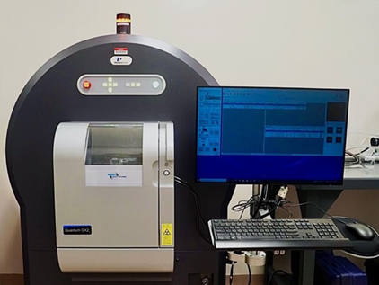
-
The TriTom Photoacoustic and Fluorescence Imaging Platform affords researchers the ability to conduct volumetric 3D imaging with the capacity for simultaneous fluorescence imaging through a tunable laser. Imaging resolutions of up to 160 x 470 micron are possible in the sagittal plane with up to 160 x 160 micron in the transverse planes allowing for close inspection whether imaging is focused on anatomical, functional, or molecular processes. In vivo imaging is performed in a temperature-controlled chamber and scan times of as little as 36 seconds allow for time effective throughput for researchers with multiple subjects. Should the focus be more aligned with contrast agent development, the cuvette sample holder can contain up to 10 contrast agents at a time to allow for rapid testing at various wavelengths.
The TriTom can be booked in 30-minute increments.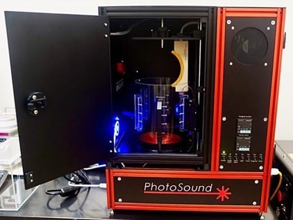
This preclinical imaging center is designed to enable true multispecies imaging, live animal imaging, and the use of state-of-the-art imaging and analysis software to offer unique insights into barriers to drug delivery, tumor biophysics, and the detection of (micro)metastases. The goal is to provide faster, higher-resolution imaging for animals and, ultimately, humans. Stefan H. Bossmann, PhD
Getting Started
All users must complete the required Animal Training through Laboratory Animal Resources Following completion of Animal Training, user training on the instrumentation can be scheduled directly with the Small Animal Imaging Center Macy Payne, PhD, facility manager. Internal users must create an iLab account for scheduling. Email Dr. Payne for instructions.
For all live animal imaging, the necessary Institutional Animal Care and Use Committee/Animal Care and Use Protocol approval must be obtained prior to initiation of any experiments. Users are asked to supply a copy of their IACUC/ACUP to Core staff prior to scheduling the first imaging session with live animals.
For all live animal imaging, the necessary Institutional Animal Care and Use Committee/Animal Care and Use Protocol approval must be obtained prior to initiation of any experiments. Users are asked to supply a copy of their IACUC/ACUP to Core staff prior to scheduling the first imaging session with live animals.
Request our Services
To learn more and utilize the Small Animal Imaging Center, contact one of its team members.Cite the cancer center support grant
This resource is funded by The University of Kansas Cancer Center Support Grant (CCSG) awarded by the National Cancer Institute (P30 CA168524). Publications that have utilized facility resources, services or scientific data generated by the resource should acknowledge the resource and cite the NCI CCSG grant.
Learn more
Interested in becoming a cancer center member?
To apply, click here. Applications are accepted throughout the year. Contact Lisa Harlan-Williams at lharlan-williams@kumc.edu for more information regarding membership.