- Home
- Types & Treatments
- Bone Cancer (Sarcoma)
- Diagnosis & Screening
Bone Cancer (Sarcoma)
Bone Cancer Screening and Diagnosis
Arriving at a diagnosis of bone cancer is a multistep process that involves:
- Possible bone cancer symptoms, such as a complaint of a mass or pain, that leads your primary care physician to conduct a physical exam. He or she may also order X-rays.
- Your primary care physician refers you to an orthopedic surgeon or specialist, who reviews the X-rays and may refer you on to an orthopedic oncologist. The orthopedic oncologist will likely order a biopsy – by needle or through surgery – or other interventional studies to help reach a diagnosis.
- A fellowship-trained specialist in the pathology of sarcoma reviews the tissue specimen collected via the biopsy and helps determine the diagnosis. This process may require 5 days or more, as bone and soft-tissue tumors are rare and require complex testing to conclusively reach a diagnosis.
How is bone cancer diagnosed?
To detect and diagnose sarcomas, our doctors perform thorough exams. They ask questions about your past health and symptoms. They will evaluate you using the latest technology. Options include:
- Computed tomography scan: A CT or CAT scan uses X-rays to make a detailed image of the structures inside the body. A contrast liquid is often injected to make the structures easier to see.
- Magnetic resonance imaging: An MRI uses a magnetic field to visualize your anatomy in a 3D format and determine the quality of the tissue.
- High-definition MRI: The University of Kansas Cancer Center offers a sophisticated, high-definition MRI. This advanced technology produces clearer images faster for a more accurate and timely diagnosis. High-definition MRI is an excellent option for people with challenging or complex conditions.
- Bone scan: This scan uses nuclear medicine to evaluate the activity of a lesion in bone.
- Positron emission tomography scan: A PET scan uses a special camera that detects cancer through the use of an injected radioactive liquid. Because cancer cells consume sugar, the accumulation of the liquid at one or multiple sites indicates cancer.
- Image-guided biopsy: Assisted by scans from an MRI and CT, we remove cells from the tumor using a small, hollow needle or through a small incision. One of our fellowship-trained specialists in the pathology of sarcoma examines the cells under a microscope to determine if they are from a sarcoma, another type of cancer or a noncancerous disease.
These tests help your healthcare team:
- Detect the presence of a sarcoma
- Pinpoint its location
- Decide what kind of sarcoma it is
- Learn how far it has spread (also called staging)
Together, this information helps your care team develop a customized treatment plan.
The University of Kansas Cancer Center offers high-definition MRI to limit the need for repeated interventional radiology. In some cases, nonsurgical cancer treatments can be image-guided to deliver the treatment directly to the affected area rather than giving the treatment systemically.

Helping others find hope
Indie rock musician Billy Brimblecom lost his left leg to bone cancer, but he didn’t let that stop him from rocking out. Now, he is on a mission to help others.
Billy’s story

Clear skies ahead
Meteorologist Gary Lezak was diagnosed with a rare tissue sarcoma. Thanks to his treatment team, he has been cancer-free since 2000.
Gary’s story
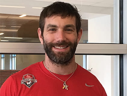
Strongman gets even stronger
Zac Craig is a Strongman competitor, athletic trainer and strength and conditioning coach. He’s also a 2-time cancer survivor.
Zac’s story
Start your path today.
Your journey to health starts here. Call 913-588-1227 or request an appointment at The University of Kansas Cancer Center.